Of course, we can not draw definitive conclusions, relying only on these parameters, as each child has his own individual schedule, depending on many factors. However, one should not neglect such important indicators. For example, according to the weight of the baby you can judge the life of the fetus, the presence of pathology, inadequate intake of nutrients or the threat of termination of pregnancy.
It is worth noting that to track how the growth and weight of the fetus varies by weeks of pregnancy, you can use ultrasound. This method allows you to get more accurate measurements of the baby. Just be sure that the baby grows and develops in accordance with the schedule can be on a routine examination, after the gynecologist measures the circumference of the abdomen and the height of the standing of the bottom of the uterus. After all, these values vary in proportion to the growth of the child for weeks of pregnancy. So, before conception, the uterus of a healthy woman of reproductive age weighs about 50-60 grams, while by the end of the period this value ranges from 1000-1300 grams. Which is quite natural, given that this body for nine months should provide the crumb comfortable conditions of life. Therefore, as the child grows, the size of the uterus increases with each week of pregnancy.
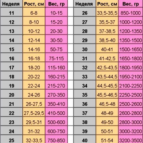
Regularities of fetal growth by weeks
There is a special table, which shows the average growth rates and weight of the fetus by week. Of course, the actual values may differ from those indicated, since these factors are influenced by various factors, including heredity. Nevertheless, in drawing up a general picture of what is happening, the correspondence of growth and weight to the norm, as well as the tendency of their increase, play an important role. As a rule, to measure the growth of the fetus begins only from the middle of the first trimester, because at the earliest dates the dimensions of the embryo are still too small.
From this point of view, it is advisable to do ultrasound before the 8th week.
At this stage, the growth of the fetus implies a distance from the crown to the tailbone. Accordingly, this size is called the coccygeal parietal and is designated only as KTP. KTP is measured up to 14-20 weeks (depending on the position of the child and the skills of a specialist who makes ultrasound) because before this time the legs of the crumb are strongly bent and it is impossible to determine the total length.
Starting from the 14-20 weeks of pregnancy, doctors try to measure the distance from the heels to the crown.
Fetal growth rates for weeks
Many women rush to make ultrasound almost immediately after the delay. In this case, the ultrasound can only confirm the presence of the fetal egg in the uterine cavity and determine its diameter. As a rule, at 6-7 midwifery week of pregnancy, this value is 2-4 mm, and on the 10th - 22 mm. Nevertheless, the future man grows intensively and develops, thus:
- by the end of 12 weeks, the CTE of the fetus is 6-7 cm;
- In the next four weeks the baby will grow 5 cm;
- and at 20 weeks the length from the crown to the heel reaches 26 cm;
- in 24 weeks - 30 cm, and in 28-35 cm;
- at 32 week, a crumb can make a jump in growth by as much as 7 cm;
- at 36 weeks most of the children are ready to be born, their organs and systems are formed, they have recovered and grown up. Basically, at this time the growth of the baby is 45-48 cm;
- nevertheless, the remaining four weeks before the cherished meeting, the crumb does not lose in vain. During this period, babies can add 100-300 grams per week and grow 1-2 cm;
- the growth of a full-term newborn baby varies within the range of 48-54 cm.