In this article, we will look at the structure of the pelvic organs of a woman, give a diagram and talk about possible anomalies in this area.
Anatomy of the pelvic organs of a woman
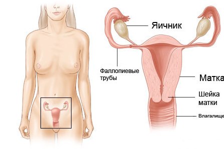
For starters, it should be noted that in the small pelvis of any person there are organs inherent in both women and men - this is the rectum and bladder. Next, we will talk about the features of the female structure of the small pelvis and those that are known only to the beautiful half of humanity.
Consider the female organs of the small pelvis on the example of the scheme:
So, this category includes fallopian tubes, uterus and cervix, as well as the vagina and the ovaries. It is these organs that are examined by a doctor of ultrasound diagnosis in case of suspicion of many diseases of the female sexual sphere, as well as when determining a possible pregnancy.
- Vagina. This organ is usually about 8 cm, it is the main participant in sexual intercourse, and in the process of childbirth becomes part of the birth canal. Inside the vagina is covered with a mucous membrane with a huge number of folds, which allows it to stretch very much to pass the newborn baby through the birth canal.
- Ovaries are responsible for the normal course of the menstrual cycle of a woman, it is in them contain eggs, and also produce female sex hormones - estrogen and progesterone. The content of these hormones in the body varies cyclically throughout life, due to which the eggs are regularly ripened. In the case of non-pregnancy, they are rejected from the body in the form of another menstruation, along with a layer of endometrium, preparing to receive a fertilized egg.
- Uterine tubes are a very important organ necessary for the conception of a future child. These tubes are sent to the uterus from the ovaries and open in its upper part. During the release of the ovum from the ovaries, the villi on the ends of the fallopian tubes can grab it and be sent to the uterus.
- The uterus is undoubtedly one of the main organs of the small pelvis in women, in appearance it resembles a pear. It is in the uterus that the fetus develops, and it grows together with its increase in size. Its walls are made up of many layers of muscles, which are rapidly stretched during the waiting period of the child. With the onset of contractions, the muscles begin to abruptly contract, thereby forcing the cervix to expand in size and open, and the fetus can enter the birth canal.
- Finally, the cervix, in fact, is its lower part, connecting the vagina and the uterine cavity.
Possible anomalies in the development of pelvic organs in women
Often during an ultrasound examination of the pelvic organs, women develop congenital malformations of the uterus, namely, a two-horned, one-horned, saddle-shaped uterus and even its bifurcation. Such features can lead to infertility, pathological miscarriage of the fetus, the threat of termination of pregnancy at any time, etc. In the case of successful childbearing, in such a situation, a planned caesarean section for the delivery of a pregnant woman is almost always scheduled.
In addition, ultrasound can also manifest acquired diseases of the pelvic organs. The most common of these are endometriosis and fibroids.
Endometriosis is a pathological process that often prevents young girls from becoming pregnant. In this disease, the endometrium grows beyond the uterine cavity, both in its walls, and into the ovaries, and even the abdominal cavity.
Myoma of the uterus, on the contrary, is usually found in women in menopause. It is a benign tumor in the female's reproductive system and requires constant monitoring in dynamics. In most cases, treatment, both in myoma and in endometriosis, is carried out in a conservative way, but only surgical surgery can completely get rid of these problems.