For one month the woman's hormonal background is very different. This is due to the preparation of her body for possible conception, and if it does not occur, the hormonal background returns to its original state. Each month, a ruptured follicle ruptures with the release of the egg, and a temporary endocrine gland, called the yellow body, is formed from the cells of the follicle itself. The role of the yellow body is to produce progesterone, which promotes the movement of the fertilized embryo into the uterus and its implantation. If conception does not occur, then the involution of the yellow body occurs after 12-14 days.
What does the yellow body look like on ultrasound?
On ultrasound, the signs of a yellow body are a non-uniform, rounded, soft-tissue sac in the ovary. If the woman has a delay in menstruation, and the yellow body is not visualized on ultrasound, the possible cause of the delay may be a disease from the endocrine or reproductive system. Even with the onset of pregnancy, the lack of visualization of the yellow body on ultrasound indicates a threat of termination of pregnancy against an insufficient level of progesterone. The dimensions of the yellow body of 18 mm are optimal for the fertilization to take place, and the embryo was implanted into the uterus and developed well. If ultrasound showed a yellow body more than 23 mm, ovulation is absent and the growth of the follicle continues, then it is called the follicular cyst. The follicular cyst can dissolve during menstruation or during the next 2-3 cycles. If ultrasound revealed a yellow body more than 30 mm in the absence of pregnancy, then it is called a yellow body cyst.
Yellow body - the size of the ultrasound
- the size of the yellow body on ultrasound 18-23 mm indicates the onset phase of ovulation and readiness for fertilization;
- the size of a yellow body on ultrasound from 20 to 30 mm during pregnancy speaks about its normal course, and in its absence about the presence of a follicular cyst;
- if during an ultrasound examination an anechogenous formation of a rounded shape larger than 40 mm in the place where the yellow body is to be found, it is customary to speak of a yellow body cyst.
Dopplerometric signs of yellow body hypofunction are found at the 13-14 week of pregnancy, when the formation of the placenta is completed, and it begins to perform the function of the yellow body for the production of progesterone.
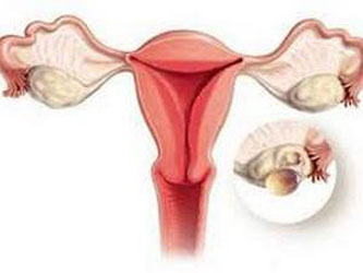
Yellow body cyst - ultrasound
As already mentioned, the yellow body of pregnancy during ultrasound is determined up to 14 weeks, and then its involution occurs. In rare cases, the extinction of the function and malnutrition of the yellow body may not occur, but its further increase and formation of a yellow body cyst, which in diameter may exceed 40 mm, occurs. This formation does not adversely affect the course and outcome of pregnancy, but with excessive growth, it is possible to compress the cyst with subsequent rupture.
A yellow body cyst can also form in the absence of pregnancy. So, 12-14 days after ovulation, in the absence of fertilization, the involution of the yellow body should occur, but if it continues to grow on the site of the burst follicle, it also leads to the formation of a yellow body cyst. In such cases, the cyst of the yellow body can be asymptomatic and be a diagnostic finding in a planned ultrasound study.
As we see, the yellow body found in the ultrasound examination of pelvic organs in women, the yellow body is an important diagnostic criterion of the reproductive function of the organism (either the ability to conceive, or the course of pregnancy in the first trimester, the threat of interruption).