What are the features of the structure of the internal genital organs?
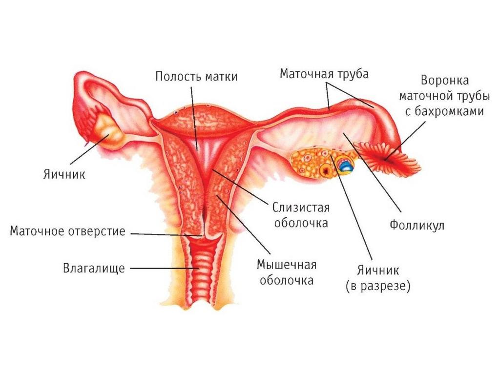
The ovary, referred to the glands of internal secretion, is a paired organ of ellipsoidal form. Its length is small - about 4 cm, and width is not more than 2.5. Despite such small size, this particular organ of the reproductive system plays the main role, synthesizing sex hormones - estrogens and progesterone.
The uterus in the anatomy of the female reproductive system, perhaps, occupies a central position. This unpaired muscular organ is the receptacle for the fetus. Despite its small size (7.5 cm in length and 5 cm in width), during pregnancy the uterus several times increases in volume and completely corresponds to the size of the fetus. This organ is located in the middle part of the pelvic cavity, directly between the bladder and rectum.
In the womb it is customary to allocate the bottom, the body, and the cervix. Normally, the cervical canal (cervical) contains mucus, which during the gestation of the child becomes denser and forms a stopper, preventing penetration of pathogens into the interior of the reproductive system.
Fallopian tubes are paired internal genital organs in women. The length of them reaches 11 cm. The uterine part (located in the wall of the uterus), the isthmus (somewhat narrowed part), the ampoule (dilated part), which ends with a funnel with numerous small outgrowths - fringes, are distinguished in each tube. It is with the help of them that there is a capture of the mature egg released into the abdominal cavity after ovulation.
The vagina is the inner sex organ in women who has direct communication with the external environment. Its length is of the order of 7-10 cm. However, in the excited state and during the course of the birth process, it can increase in size. This is due to the smoothing of the inner folds of the organ.
What are the characteristics of the structure of the external genitalia in women?
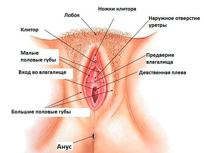
In order to fully understand how the female reproductive system is arranged, let us consider those anatomical entities that are referred to the external genitalia.
The pubis is a part of the lower part of the anterior abdominal wall, which has a triangular shape and when covered with puberty, it is covered with hair. It is located directly in front of the lone articulation. Has a well-pronounced subcutaneous fat.
From below the pubis turns into the large labia, - paired, round folds of about 7 cm in length, and not more than 2 cm in width. The skin of the outer surface of the lips is covered with hair. In the thickness of this anatomical formation is located subcutaneous fatty tissue.
Small labia hide behind large ones and are nothing more than skin folds. In front, they are connected by a soldering, which covers the clitoris, and behind it merge into the rear soldering.
The clitoris is similar in its internal arrangement to the male penis. It consists of cavernous bodies that fill with blood during intercourse and increase the size of the body.
The hymen is a thin mucous membrane that covers the entrance to the vagina. During the first sexual intercourse, it ruptures, which is accompanied by a slight bleeding.
Bartholin glands are located in the thickness of the large labia. During sexual intercourse, they are secreted lubrication, which moistens the vagina.
In order to better imagine the structure of the female reproductive system, of what it consists of, we will provide a diagram, which clearly shows the location of its main organs.