Trisomy on the 21st chromosome, or Down's Syndrome, is the most common form of genomic pathology that occurs in about 1 out of 800 children born. Scientists found that the disease is due to an incorrect distribution of chromosomes, resulting in the patient, instead of the two copies of the 21st chromosome, there are three. To anticipate the appearance of pathology is impossible, it is obvious that one-trisomy on the 21st chromosome means nothing other than a series of mental, physical and behavioral disorders that interfere with the normal development and existence of a sick child.
In connection with the above, it is difficult to overestimate the importance of prenatal diagnosis, allowing in utero to determine the risk of trisomy 21 by characteristic indicators.
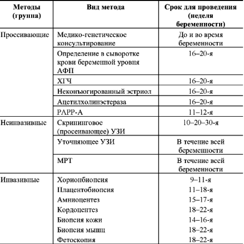
First trimester screening
Refers to non-invasive methods and consists of ultrasound and a biochemical analysis of the mother's blood. The optimal time for the first prenatal screening is 12-13 weeks.
During ultrasound diagnosis, specialists pay attention to the size of the collar zone, which is a characteristic marker of the presence of abnormalities. Namely, depending on which week of pregnancy and the norm corresponding to it, the sign of trisomy 21 may be the expansion of the collar space by more than 5 mm.
In turn, the woman's blood is examined for two hormones: free b-HCG and RARR-A. For the unit of measurement of the studied indicators take - MoM. The values obtained are compared with the normal values: Trisomy 21 may indicate an increased level of free b-hCG - more than 2 M0Ma, and the concentration of PAPP-A is less than 0.5MoM.
However, based on the results of the first prenatal screening, it is impossible to draw definitive conclusions, because this is only a probabilistic indicator that does not always take into account other factors influencing the level of these hormones. To them it is possible to carry: incorrectly specified term of pregnancy, weight of mother, stimulation of ovulation, smoking.
Second prenatal screening
In the interval between 15-20 weeks, a second attempt is made to diagnose genomic pathology. This period is considered more informative, because many violations can be seen during the ultrasound. For example, in a fetus with trisomy on 21 chromosomes differ from the norm: the length of the humerus and femur, the size of the bridge of the nose, the size of the renal pelvis, and sometimes the visual defects of the heart, gastrointestinal tract or cyst of the vascular plexus of the brain.
Blood of a pregnant woman is examined for AFP level, which is a bright marker of the hereditary pathology of the fetus. If, as a result of the second screening, the AFP was found to be below normal, then this could indicate the presence of trisomy on 21 chromosomes.
The results obtained are compared with the results of the first study, if the risks are sufficiently high, the pregnant woman is assigned other methods of examination.
Invasive methods for determining chromosomal abnormalities
More accurate, but also more dangerous ways to determine genomic disorders are:
- amniocentesis - means piercing of the abdominal wall with a thin needle and fence of the amniotic fluid, which is investigated in the future;
- Chorion biopsy is a fairly informative method. As a rule, sampling of villi and placenta cells is performed at 11-12 weeks, by piercing the abdominal cavity, or by using a catheter through the vagina;
- umbilical cord blood sampling, is performed no earlier than the 18th week of pregnancy.
Invasive methods, although they allow more accurate determination of the presence of a genomic anomaly, but at the same time carry the risk of arbitrary termination of pregnancy.